Dental X-rays are safe when a dentist orders them for a specific clinical reason and takes standard dose-reduction steps. The ADA’s current recommendations confirm this, built around the ALARA and ALADA principles: as low as reasonably achievable, and as low as diagnostically acceptable. The one exception worth flagging is CBCT, or cone beam CT, which delivers a meaningfully higher dose and should only be used when a flat 2D image genuinely will not answer the clinical question.
TL;DR:
- Modern digital equipment and proper technique significantly reduce radiation doses, making routine intraoral X-rays comparable to a few days of natural background radiation.
- CBCT scans deliver higher doses depending on the field of view, so they should only be used when detailed 3D imaging will truly impact diagnosis or treatment planning.
- Dental X-ray radiation’s link to cancer is inconclusive, but the small risk is outweighed by the benefits of diagnosing conditions like infection or decay early.
- The necessity of X-rays depends on clinical needs, with imaging recommended only when it will directly influence diagnosis or treatment, not on a fixed schedule.
- Best practices include asking why an X-ray is ordered, whether prior images exist, and limiting CBCT use to cases with clear justification.
questions about this? we're happy to help.
Every patient's situation is a little different. If you want to talk through what this means for you, our Greenville team is one call away.
schedule a free consultationTable of Contents
- What Are the Different Types of Dental X-Rays?
- How Much Radiation Do Dental X-Rays Actually Deliver?
- Does Dental X-Ray Radiation Cause Cancer?
- When Are Dental X-Rays Actually Necessary?
- How Do Dentists Minimize Radiation Exposure?
- Are Dental X-Rays Safe During Pregnancy and for Children?
- Should You Decline Dental X-Rays, and How Often Do You Really Need Them?
- How Complete Dental Care Applies This Guidance in Practice
- What Happened to Lead Aprons and Thyroid Collars?
- What Regulations Govern Dental X-Ray Equipment?
- Why Does Staff Training Matter as Much as the Equipment?
- How Are X-Ray Materials and Equipment Disposed Of Safely?
- How Do Dentists Explain X-Ray Risks and Benefits to Patients?
- What Actually Matters Here, and What Gets Overblown
- Exams, X-Rays, and Straight Answers at Complete Dental Care
- Sources
- FAQ
What Are the Different Types of Dental X-Rays?
Dental imaging splits into two broad categories, and the difference matters for understanding dose. Intraoral X-rays place the sensor or film inside the mouth and capture a narrow, detailed view of a few teeth. Extraoral X-rays sit outside the mouth and capture a wider picture of the jaw, skull, or full arch.
Each type has a specific job:
- Bitewing X-rays catch the crowns of upper and lower back teeth in one image, mainly to spot cavities between teeth and check bone height at the gumline.
- Periapical X-rays show a full tooth from crown to root tip, used when a dentist needs to check for infection, abscess, or bone loss around one specific tooth.
- Panoramic X-rays sweep the entire jaw, sinuses, and joint areas in a single flat image, useful for planning orthodontics, checking wisdom teeth, or getting a broad baseline view.
- CBCT (cone beam computed tomography) builds a 3D volume of the jaw, teeth, and surrounding bone, typically reserved for implant planning, complex root canal anatomy, or surgical cases where depth and precise measurements matter.
That last category is where the safety conversation gets more nuanced. CBCT gives a dentist information a flat image simply cannot: precise bone volume before placing an implant, or the exact path of a curved root canal. But that extra detail comes from a scan that irradiates a larger volume of tissue, which is why professional guidance treats it as a tool for specific cases, not a routine upgrade.
How Much Radiation Do Dental X-Rays Actually Deliver?
A single bitewing X-ray delivers a fraction of the radiation most people absorb from the environment in an ordinary day. That comparison is the fastest way to put dental imaging dose into perspective, and it’s the one dentists reach for most often when a nervous patient asks the obvious question.
The NRC’s dose reference data puts the average American’s annual background radiation exposure, from soil, cosmic rays, radon, and the food supply, in a range most people never think about. Dental X-rays add a small increment on top of that baseline:
- Bitewing and periapical X-rays deliver the lowest doses of any dental image, comparable to a few hours to about a day of ordinary background exposure.
- Panoramic X-rays run somewhat higher than a single bitewing, since they capture the entire jaw in one pass, but still sit well below a medical chest X-ray in most comparisons.
- CBCT scans deliver the highest dose among dental images, with a range that varies significantly based on field of view. A small, focused scan for one implant site costs far less than a large scan covering both jaws.
A dental bitewing typically delivers roughly the same radiation dose as spending a single day exposed to natural background radiation. That comparison alone explains why panels like the ADA treat routine intraoral imaging as low risk when clinically justified.
The wide range in CBCT dose numbers is not a data problem. It reflects real variation in scanner settings, field of view, and exposure time between machines and practices. A dentist scanning one tooth for a root canal uses a fraction of the radiation of a full-arch scan for complex implant planning, which is exactly why field-of-view selection matters as much as the decision to use CBCT at all. Chest X-rays, for comparison, remain a useful benchmark because most patients have had one and can mentally calibrate against it. Nearly every category of standard dental imaging falls below that mark, with CBCT being the notable exception depending on scan size.
Does Dental X-Ray Radiation Cause Cancer?
The honest answer is that the evidence is mixed, and the absolute risk described in the research is small. A peer-reviewed review published on PMC examined the body of research on dental diagnostic X-ray exposure and found that some individual studies suggested possible associations with certain cancers, including meningioma and thyroid cancer. The same review is clear that this evidence is limited and inconsistent across studies, not a settled causal finding.
That distinction between “some studies suggest an association” and “X-rays cause cancer” is where a lot of anxious internet searching goes wrong. Here’s why the science is genuinely hard to pin down:
- Low-dose epidemiology is difficult by nature. When the radiation dose per exposure is small, detecting a cancer signal requires enormous study populations followed over decades, and confounding factors (other radiation sources, genetics, unrelated risk factors) are hard to fully separate out.
- Recall bias affects older studies. Many of the studies examining past dental X-ray exposure and cancer relied on patients or family members recalling how many X-rays they had decades earlier, an approach that introduces real uncertainty into the data.
- Imaging technology has changed dramatically. Older studies often reflect film-based X-ray equipment from decades past, which used considerably higher doses than the digital sensors and collimated beams standard in dental offices today.
- Study designs vary widely. Case-control studies, which compare people who already have a disease to those who don’t, are more prone to bias than the large prospective studies that would give more definitive answers, and prospective studies on dental X-ray exposure specifically are scarce.
None of that means concerns about the associations found in some studies should be waved away. It means the evidence supports a measured position: the absolute risk from properly justified, modern dental imaging is very small, while the risk from skipping necessary imaging (a missed abscess, an unnoticed area of bone loss, an undetected fracture) is often more immediate and concrete. That’s the calculation a dentist is actually making every time an X-ray gets ordered, and it’s why “only when clinically necessary” isn’t a throwaway phrase. It’s the entire operating principle behind current guidance.
When Are Dental X-Rays Actually Necessary?
The ADA’s updated recommendations state plainly that dental X-rays should be ordered only when the images will actually affect diagnosis or treatment planning, not on a fixed calendar schedule applied to every patient regardless of risk. That’s a shift away from the old assumption that everyone needs bitewings once a year no matter what.
Dentists weigh several factors before deciding an X-ray is warranted:
- Current symptoms. Pain, swelling, a broken tooth, or a suspicious spot on the gums all point toward imaging that addresses that specific complaint.
- Availability of prior images. If a dentist can pull recent, diagnostic-quality X-rays from a previous provider, a repeat exposure often isn’t needed.
- Caries risk level. A patient with a history of frequent cavities and poor enamel may need more frequent bitewings than someone with a clean record and good home care.
- Treatment planning needs. Implant placement, orthodontic work, or root canal therapy each carry their own imaging requirements, sometimes including CBCT when the case calls for 3D detail.
- Age and developmental stage. Growing jaws and emerging teeth in children sometimes warrant imaging that wouldn’t apply to an adult with a stable, fully erupted bite.
Before agreeing to any X-ray, it’s reasonable to ask a few direct questions: What specifically are you looking for? Do you have my prior images on file? Would a lower-dose image work instead of CBCT? How will this change my treatment plan?
Pro Tip: Ask your dentist’s office to request your X-rays from a previous practice before your first visit. Most offices can transfer digital images electronically in a day or two, which often eliminates the need for a full new set at your first appointment.
How Do Dentists Minimize Radiation Exposure?
Modern dental offices lean on two guiding principles: ALARA (as low as reasonably achievable) and ALADA (as low as diagnostically acceptable). The ADA describes ALADA as the practical extension of ALARA for imaging specifically, meaning the image chosen has to be good enough to answer the clinical question without reaching for a higher-dose option than the situation requires.
In practice, that translates into specific equipment and technique choices, not just a general philosophy:
- Digital sensors replace older film, requiring a significantly lower radiation dose to produce a usable image while also letting dentists enhance and zoom digitally instead of retaking a shot.
- Rectangular collimation shapes the X-ray beam to match the size of the sensor, cutting the amount of surrounding tissue exposed compared to older round-beam collimators.
- Receptor holders keep the sensor stable and correctly positioned, reducing retakes caused by blurry or poorly angled images.
- Longer source-to-skin distance spreads the beam out before it reaches the patient, lowering the dose at the skin surface.
- Technique charts standardize exposure settings by patient size and image type, a straightforward control that FDA technical guidance flags as an underused way to cut dose when staff actually follow it consistently.
Equipment alone doesn’t guarantee safety. Staff training and routine quality assurance testing on X-ray machines matter just as much, since a well-designed sensor paired with outdated technique settings or an uncalibrated machine still delivers more radiation than necessary.
Pro Tip: If you’re curious whether a practice follows modern protocols, ask whether they use digital sensors and rectangular collimation. Both are now standard in offices that have updated their equipment in the last several years, and most staff will answer the question readily.
Are Dental X-Rays Safe During Pregnancy and for Children?
Pregnancy tends to be the single biggest source of dental X-ray anxiety, and the guidance here has shifted in a direction that surprises a lot of patients. Current ADA guidance notes that lead abdominal aprons and thyroid collars are no longer generally recommended as a routine step, because modern beam collimation and digital receptors already keep radiation tightly focused on the target area. Necessary dental X-rays, taken with proper technique, are considered safe during pregnancy when a real clinical need exists, such as diagnosing an infection that could otherwise threaten the pregnancy if left untreated.
For children, the guiding idea is “child-size” exposure. Pediatric dentistry guidance from the AAPD calls for individualized imaging decisions based on each child’s caries risk and developmental stage, not a blanket schedule applied to every young patient. Children’s exposure settings should be adjusted for their smaller size, and CBCT use in kids should be limited to cases where the diagnostic benefit clearly outweighs a routine 2D image.
A few practical points apply across both groups:
- Pregnant patients should tell their dentist before any imaging is scheduled, even though shielding recommendations have changed.
- Parents can ask whether a pediatric-specific exposure setting is being used for their child’s X-ray.
- Prior images should travel with the patient between providers whenever possible, since reviewing existing images before ordering new ones is one of the simplest ways to avoid an unnecessary repeat exposure.
Should You Decline Dental X-Rays, and How Often Do You Really Need Them?
Declining an X-ray is a reasonable choice in some situations, but it comes with trade-offs worth understanding before you say no. If a dentist can’t visually or clinically justify a need, or if you have recent diagnostic-quality images available, pushing back is fair. If a dentist genuinely can’t assess a hidden problem like decay between teeth or infection at a root tip without imaging, declining means that condition could go undiagnosed until it becomes more serious and more expensive to treat.
A few things to know before your next appointment:
- You can ask for the clinical reason behind every X-ray order, not just accept it as routine paperwork.
- Frequency should follow your individual risk, not a fixed calendar. Someone with a clean cavity history and healthy gums may go longer between bitewings than someone with active decay.
- You can request that your dentist document a refusal in your chart rather than simply skip the conversation, which protects both of you if a condition is later discovered.
- CBCT should come with a specific justification, such as implant planning or complex root canal anatomy, not a general “let’s get a better look” without a defined clinical question.
- Bringing prior images to a new dentist is one of the most effective ways to avoid a repeat scan you don’t actually need.
How Complete Dental Care Applies This Guidance in Practice
At Complete Dental Care, imaging decisions follow the same ALARA and ALADA principles outlined in current ADA guidance, not a fixed once-a-year routine applied to every patient regardless of history. That means digital sensors and rectangular collimation are standard for intraoral imaging, and CBCT gets reserved for cases where it genuinely changes the plan, like implant placement or a root canal with unusual anatomy.
In practice, that looks like a few specific habits. New patients are asked about recent X-rays from a previous dentist before ordering new ones. Pregnancy is flagged at intake so imaging decisions and technique choices reflect that conversation from the start. Treatment planning for implant work can rely on 3D imaging because that level of detail supports accurate implant placement.
If you’re weighing whether an X-ray is necessary for your situation, ask us directly. We’ll walk through what we’re looking for and why, and if you have images from another provider, we’d rather see those first than order a repeat scan you don’t need.
What Happened to Lead Aprons and Thyroid Collars?
If you remember being draped in a heavy lead apron before every dental X-ray, the newer approach might catch you off guard. The ADA’s current position is that routine lead abdominal aprons and thyroid collars are no longer generally recommended for most intraoral X-rays, a genuine reversal from decades of standard practice.
The reasoning comes down to how modern equipment controls the beam. Rectangular collimation, tighter beam limitation, and digital receptors already confine radiation closely to the target area, which makes external shielding largely redundant for routine images. There’s also a practical downside to shielding that rarely got mentioned in the past: a poorly positioned lead apron can block part of the primary beam, forcing a retake, which means the patient ends up exposed twice instead of once. Recent recommendation changes prioritize getting a usable image on the first try over layering on a shielding step that no longer adds meaningful protection with today’s equipment.
This doesn’t mean shielding is banned or wrong to ask about. Some states and practices still follow local regulations that require it, and patients who feel more comfortable with a lead apron can generally still request one. The larger point is that the safety of the image now rests primarily on beam control and technique, not on layers of external shielding, which is a genuine shift in how the profession thinks about protecting patients.
What Regulations Govern Dental X-Ray Equipment?
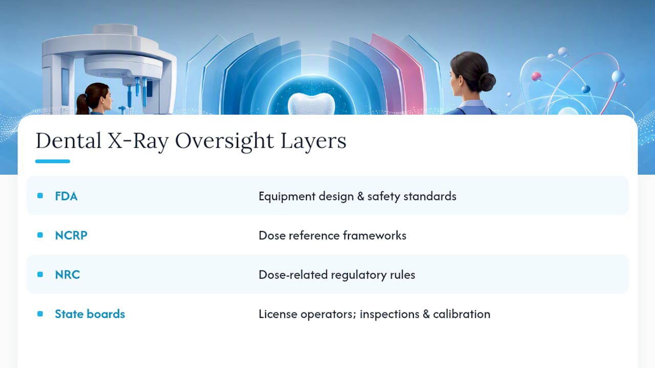
Dental X-ray machines and the people operating them fall under a layered system of federal and professional oversight, not a single agency’s rulebook. The FDA regulates the equipment itself, setting design and performance standards manufacturers must meet before a machine reaches a dental office, including specific safety considerations for hand-held X-ray units that carry unique exposure risks for the operator holding the device.
The National Council on Radiation Protection and Measurements (NCRP) and the Nuclear Regulatory Commission (NRC) provide dose reference frameworks that inform how radiation exposure limits and safety benchmarks get set across medical and dental imaging. State dental boards typically layer additional requirements on top of federal rules, covering who is licensed to operate imaging equipment and how often machines must be inspected and calibrated.
None of this operates as background paperwork patients never see the effects of. It’s the reason a dental X-ray machine gets periodically tested for output accuracy, why operators need documented training before they can position and fire the equipment, and why a hand-held unit used for house calls or mobile dentistry has to meet specific FDA safety criteria different from a wall-mounted unit bolted into an exam room. Patients rarely ask about equipment calibration schedules, but the answer to “when was this machine last tested” says a lot about whether a practice takes the regulatory framework seriously or treats it as a checkbox.
Why Does Staff Training Matter as Much as the Equipment?
A dental office can own the newest digital sensor on the market and still deliver more radiation than necessary if the person operating it hasn’t been properly trained on positioning, exposure settings, and when to stop and consult rather than retake a poor image. Technique charts, the size-based exposure protocols mentioned in FDA technical guidance, only reduce dose when staff actually reference and follow them consistently, rather than defaulting to one exposure setting for every patient regardless of size or age.
Quality assurance programs close that gap. They typically include periodic equipment testing to confirm output matches the manufacturer’s specifications, documented staff competency checks, and a process for reviewing retake rates, since a high number of repeat images often signals a training gap rather than a hardware problem. Retakes matter for a simple reason: every repeated image doubles that patient’s exposure for no diagnostic gain.
Training also covers judgment calls that don’t show up in a technical manual. Knowing when a CBCT field-of-view should be sized down for a single implant site instead of scanning the full jaw, or recognizing when a finding on a scan exceeds what a general dentist should interpret alone, is where the AAPD’s guidance on CBCT interpretation points toward consulting a radiologist rather than guessing. That kind of institutional discipline, built through ongoing training rather than a one-time certification, is a quieter part of dental X-ray safety than the equipment itself, but it’s arguably just as important.
How Are X-Ray Materials and Equipment Disposed Of Safely?
Dental X-ray equipment and the materials associated with it carry environmental handling requirements that mostly happen behind the scenes, but they matter for anyone curious about the full safety picture. Older lead aprons and thyroid collars, now less commonly used given the shift in shielding recommendations, still require specific disposal or recycling processes because lead is a regulated hazardous material that can’t simply go into standard office trash.
Digital sensors and the computer hardware that supports them fall under electronic waste guidelines when a practice upgrades equipment, since circuit boards and imaging sensors contain materials that shouldn’t go to a landfill. Older X-ray machines themselves, when decommissioned, typically require disposal through vendors equipped to handle the internal components safely, particularly any residual lead shielding built into the machine housing.
Film-based X-ray processing, largely phased out in favor of digital sensors, historically involved chemical developer and fixer solutions that required regulated disposal due to their silver content, one more reason the shift toward all-digital imaging has had a genuine environmental upside beyond the dose reduction for patients. A practice that has fully transitioned to digital sensors has, in effect, eliminated an entire category of hazardous chemical waste that used to be routine in every dental office.
How Do Dentists Explain X-Ray Risks and Benefits to Patients?
The best conversations about dental X-rays happen before the image is taken, not after a patient asks a worried question in the parking lot. A clear explanation typically covers three things: what specifically the dentist is looking for, why a visual exam alone can’t answer that question, and what the radiation dose looks like in relatable terms, often by comparing it to a day or two of ordinary background exposure.
Good patient communication also means being upfront about uncertainty rather than offering false confidence in either direction. Telling a patient “X-rays carry zero risk” overstates the case the PMC review actually supports, while implying a scan is dangerous when it’s clinically necessary can lead a patient to decline imaging that would have caught a real problem early. The more honest framing acknowledges a small, well-studied risk exists alongside a clear diagnostic benefit, and lets the patient weigh that trade-off with actual information instead of guesswork.
Dentists who do this well also invite questions rather than waiting for them. Explaining why a CBCT scan is being ordered instead of a standard panoramic image, describing what happens to prior images requested from another office, or simply asking a pregnant patient whether she has any concerns before positioning the sensor, all signal that the imaging decision is being made with the patient rather than around them.
What Actually Matters Here, and What Gets Overblown
The conventional advice on dental X-ray safety tends to fixate on the wrong variable. Patients worry most about the moment of exposure itself, the beep of the machine, the sensation of biting down on a sensor, when the more consequential decision happened earlier: whether that X-ray was clinically justified in the first place. A properly justified, modern digital X-ray carries a small, well-documented dose. An unjustified one, however low the dose, is radiation exposure with no offsetting benefit, and that’s the real distinction worth caring about.
The shift away from routine lead aprons is the clearest sign that this field updates its thinking when the evidence changes, even when that update contradicts what patients have been told for decades. That should build confidence in the process, not undermine it. What we’d urge readers to prioritize first is simple: ask why an X-ray is being ordered, ask whether prior images exist, and treat CBCT as a scan that needs its own specific justification, not an automatic upgrade. Those three habits do more for your safety than worrying about the dose itself.
— Complete Dental Care
Exams, X-Rays, and Straight Answers at Complete Dental Care
Complete Dental Care treats imaging as a diagnostic decision, not a routine line item, which means you get a clear reason for every X-ray rather than a scan added by default. Our Exams & X-Rays visits use digital sensors and beam collimation as standard, and we’ll walk you through why a particular image is needed before it’s taken. If you’re pregnant or have concerns about imaging during pregnancy, our X-ray safety during pregnancy page covers exactly how we handle those appointments. Considering full-arch implants or another procedure where 3D imaging plays a role in planning? Our surgical and implant dentistry page explains how that imaging fits into the treatment process. Have a specific question about your own X-ray history or an upcoming appointment? Contact our office and we’ll talk it through before you’re in the chair.
This article is general information, not a substitute for advice from a qualified doctor. Consult a qualified healthcare professional about your own circumstances before acting on anything here.
Sources
- New Recommendations Confirm Dental X-Rays Most Effectively Used in Moderation | American Dental Association
- Health effects from exposure to dental diagnostic X-ray
- Doses in Our Daily Lives | U.S. Nuclear Regulatory Commission (NRC)
FAQ
Should I decline dental X-rays?
Declining is reasonable when a dentist can’t give a specific clinical reason for the image or when recent diagnostic-quality images already exist. If a dentist needs to check for hidden decay, infection, or bone loss that a visual exam can’t reveal, declining means that condition could go undetected until it’s more advanced.
Are dental X-rays safe now?
Yes, when ordered for a clear clinical reason and taken with modern digital equipment and beam collimation, following the ADA’s current recommendations. The main exception is CBCT, which carries a higher dose and should be reserved for cases where standard imaging isn’t enough.
Is radiation exposure from dental X-rays harmful?
The radiation from routine bitewing and periapical X-rays is very low, often comparable to a day or so of ordinary background exposure from the environment. A peer-reviewed review found some studies suggesting possible associations with certain cancers, but described the evidence as limited and inconsistent rather than conclusive.
Is it really necessary to get X-rays at the dentist?
X-rays are necessary when a dentist has a specific diagnostic question that a visual exam can’t answer, such as decay between teeth or infection at a tooth root. Current guidance recommends imaging only when it will affect diagnosis or treatment, not on a fixed yearly schedule for every patient.
How often should dental X-rays be taken?
Frequency should be based on individual risk factors like cavity history, gum health, and prior treatment needs, not a routine calendar interval applied to everyone. Someone with a clean history and low caries risk may go longer between imaging than someone with active dental problems.
questions about this? we're happy to help.
Every patient's situation is a little different. If you want to talk through what this means for you, our Greenville team is one call away.
schedule a free consultation